Xerophthalmia

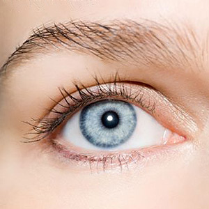
Xerophthalmia is commonly known as Dry Eye Syndrome. It is a caused by a severe drying of the eye surface or the conjunctiva due to the inability of the lacrimal or tear glands to produce tears. This chronic autoimmune disease results in dryness, ulceration and keratomalacia which is a softening of the cornea leading to blindness. It is the most common and yet preventable cause of blindness over the world and has been a main cause of visual disability among young children in many places.
It occurs in infants when they are weaned off food rich in Vitamin A or when young children do not eat many vegetables. It is also common among the older population, and those who suffer from autoimmune disorders such as such as lupus or Sjögren's syndrome. However the peak incidence is noticed in children between the ages of 3 to 5 years. It usually occurs in otherwise healthy individuals.
Causes of Xerophthalmia
Xerophthalmia is often associated with systemic diseases such as Sjogren' s syndrome, systemic lupus
erythematosus, rheumatoid arthritis, amyloidosis, hypothyroidism; deficiency of vitamin A, usage of medications such as antihistamines, nasal decongestants, tranquilizers, and anti-depressant drugs. Thermal or chemical burns can also cause the disease.
The abnormal dryness and thickening of the conjunctiva and cornea may be caused due to trauma, local disease, conditions in which the eyelids do not close completely or systemic deficiency of Vitamin A due to an inadequate diet. This systemic deficiency of Vitamin A may occur due to absorption problems, deficiency of protein or zinc which can prevent the release of Vitamin A from the liver, insufficient number of calories, problems with the conversion of beta carotene to retinol and Vitamin A storage problems.
Symptoms of Xerophthalmia
The earliest symptoms include feeling as though there is a foreign object in the eye and mild irritation.
There is a feeling of hot discomfort and a desire to blink continuously. Later on, there is severe sensitivity to light. The symptoms of the disease include night blindness and appearance of Bitot's spots on the conjunctiva.
A decrease in visual acuity is noticed. These are small plaques which are grey in color, triangular or oval in shape and can be removed by administration of Vitamin A doses. However they also appear in healthy individuals. Till this stage the condition is reversible. Photophobia may occur and dull hair and dry mouth are commonly seen.
The white portion of the eye becomes dry and wrinkled. Loss of bone, tooth enamel, and taste and smell are other symptoms. Diarrhea, respiratory infections and reduced immunity may also be seen. Eye and eyelid movements remain normal. Salivation, hearing and sense of balance all remain intact. Reduced immunity makes the patients more vulnerable to infections, especially in infants and children.
Diagnosis of Xerophthalmia
The 'Tear film break up line' is used to clinically diagnose the disorder. It is the interval between a complete blink and the appearance of the first dry spot on the cornea. This is supposed to occur between 15 to 30 seconds. If it occurs within 10 seconds, it indicates severe deficiency. The Schirmer's test is performed by inserting paper strips or filter paper into the eye for a number of minutes to check whether enough tears are being produced
Treatment of Xerophthalmia
Commencing treatment and making the appropriate changes in lifestyle and diet as soon as possible may improve the scope for recovery. An ophthalmologist can detect this by performing an eye examination. Medications such as Restasis, topical corticosteroids, and oral tetracycline and doxycycline may be recommended by the doctor. Most patients with this disorder only experience discomfort and no vision loss.
As previously mentioned, Vitamin A doses may be given to correct the deficiency and cause the eye to heal. However, these take several days to show their effect and at least three doses are required. Antibiotic ointments, hydrophilic contact lenses and eye drops are used to control infection and keep the eye surface moist. Tear substitutes or artificial tears which are available over the counter are also used. In severe cases the duct that drains away the tears from the eye is permanently closed.
As diet is an important contributing factor, the patients are advised to increase their intake of green leafy vegetables and milk. A protein rich diet is also recommended. Vitamin A or Beta Carotene ought to be increase in the diet. Children with severe protein malnutrition must be referred to a hospital and carefully monitored. They must avoid heat, wind and strong light. Smoke filled rooms and straining the eye must be avoided. Wearing wraparound glasses and using humidifiers to maintain the humidity in the air can relieve discomfort.
Women of reproductive age with Bitot's spots or night blindness must be given a daily oral dose not exceeding 5,000-10,000 IU of vitamin A for at least 4 weeks. They must be careful not to exceed this limit namely 3000 micrograms as it may cause birth defects. The three dose treatment must also be administered.
Drinking carrot juice and intake of two tablespoons of cod liver oil everyday can help alleviate some of the symptoms. Dark green, yellow and orange colored vegetables, fish liver, beef, pork and other livers, mango, papaya, eggs, butter, broccoli and apricots are among the recommended dietary guidelines. Slow neck rolls and other eye exercises should be performed and it is important to remain optimistic. Smoking and exposure to secondary smoke must be avoided.
Top of the Page: Xerophthalmia
Tags:#xerophthalmia #xerophthalmia symptoms #xerophthalmia causes #xerophthalmia treatment #dry eye syndrome
Stress and Brain Damage
Lewy Body Dementia
Pervasive Development Disorders
Sinus Infection
Computer Vision Syndrome
Lasik Eye Surgery
Xerophthalmia
Dry Eye Syndrome
Peripheral Neuropathy
Cerebral Palsy
Migraine Headache Symptom
Low Blood Sugar Headache
Dizzy Spells
Tinnitus
Oral Hygiene
Halitosis
Treatment for Gingivitis
Neck Pain Causes
Whiplash Injury
Other health topics in TargetWoman Women Health section:
General Women Health

Women Health Tips - Women Health - key to understanding your health ...
Cardiac Care
Women's Heart Attack Symptoms - Identify heart problems...
Skin Diseases
Stress Hives - Red itchy spots ...
Women Disorders
Endocrine Disorder - Play a key role in overall wellbeing ...
Women's Reproductive Health
Testosterone Cream for Women - Hormone replacement option ...
Pregnancy
Pregnancy - Regulate your lifestyle to accommodate the needs of pregnancy ...
Head and Face
Sinus Infection - Nearly 1 of every 7 Americans suffer from ....
Women and Bone Care

Slipped Disc - Prevent injury, reduce pain ...
Menstrual Disorders
Enlarged Uterus - Uterus larger than normal size ...
Female Urinary Problems
Bladder Problems in Women - Treatable and curable ...
Gastrointestinal Disorders
Causes of Stomach Ulcers - Burning feeling in the gut ...
Respiratory Disorders
Lung function Test - How well do you breathe ...
Sleep Management

Insomnia and Weight Gain - Sleep it off ...
Psychological Disorders in Women
Mood swings and women - Not going crazy ...
Supplements for Women
Women's Vitamins - Wellness needs...
Natural Remedies

Natural Diuretic - Flush out toxins ...
Alternative Therapy
Acupuncture Point - Feel the pins and needles ...
Top of the Page: Xerophthalmia
Popularity Index: 101,482

